Many of the contraction principles that apply to skeletal muscle also apply to smooth muscle. Most importantly, essentially the same attractive forces that occur between myosin and actin fibers in skeletal muscle also cause smooth muscle contraction, but the internal physical regulation of actin and myosin filaments in smooth muscle fiber differs from skeletal muscle.
Smooth muscle types
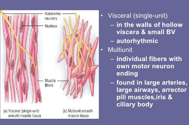
In general, smooth muscle can be divided into two main classes:
• Multi-unit smooth muscle. The most important feature of multiunit smooth muscle fibers is that each glass fiber can contract independently of the others and is regulated more often by nerve signals. Examples include ocular smooth muscle fibers, iris, and piloerector muscles that cause hair loss when activated by the sympathetic nervous system. • Individual smooth muscle. This type is also called unit smooth muscle, syncytial smooth muscle, and visceral smooth muscle. The mass of hundreds to millions of muscle fibers shrinks as a whole. Cell membranes are joined in gap junctions, so action potentials can travel from one fiber to another and cause muscle fibers. This type of muscle is found in the walls of the digestive tract, bile ducts, ureters, uterus, fallopian tubes and blood vessels.
Physical Basis for Smooth Muscle Contraction
Smooth muscle does not have the same radiant arrangement of actin and myosin filaments as skeletal muscle
• Actin fibers adhere to dense bodies. Some of the dense bodies are scattered in the cell and are bounded by a skeleton of protein structures that connect one dense body to another. Some adhere to the cell membrane and form bonds with the dense bodies of neighboring cells, allowing the transfer of holding force from one cell to another.
• Myosin fibers are inserted between actin fibers. Myosin fibers are more than twice the diameter of actin fibers. • Contractile units. The individual contractile units consist of actin fibers radiating from two dense bodies; these fibers overlap the myosin fiber located in the middle of the dense bodies.
Comparison of Smooth Muscle Contraction and
Skeletal Muscle Contraction
Unlike skeletal muscle contractions, most smooth muscle contractions last for several hours or even days of tonic. The physical and chemical properties of smooth muscle differ from those of skeletal muscle. Here are some of the differences:
• Slow driving on cross bridges. The rate of cruciate ligament cycling in smooth muscle (i.e., the rate of attachment and release of myosin cruciate ligament with actin) is slower in smooth muscle than in skeletal muscle.
• Low power consumption. Only 1/10 to 1/300 of the force is needed to maintain a smooth muscle contract compared to skeletal muscle.
• Slow onset of shrinkage and relaxation. Normal smooth muscle tissue begins to contract 50 to 100 milliseconds after lifting and has a total retention time of 1 to 3 seconds, which is 30 times longer than normal skull muscles.
• Increased maximum shrinkage force. The maximum contractile force of smooth muscle is always greater than that of skeletal muscle. This extra force of attraction is postulated as a result of the long binding time of myosin crosslinks to actin fibers.
Smooth muscle can be shortened by a greater percentage of its length than cranial muscle. Skeletal muscle has a useful reduction distance of about one-third to one-third of its extended length, while smooth muscle often contracts and is more than two-thirds of its extended length.
The "latch mechanism" facilitates long-term holding of contractions. Once a smooth muscle develops a full contraction, the level of muscle activation usually decreases to a level lower than the first level, but the muscle can maintain its full contractile force. This is called a "locking mechanism". The importance of the latching mechanism lies in the fact that it can maintain a longer tonic contraction of smooth muscles for hours with minimal energy consumption.
Regulation of calcium ion contraction
Calcium ions combine with calmodulin to stimulate myosin kinase activation and myosin head phosphorylation. Smooth muscle lacks troponin, but instead contains calmodulin, another regulatory protein. Although this protein reacts with calcium ions, it differs from troponin in the way the contract begins; This is done by calmodulin activation of myosin crosslinks. Regulation of contractions is myosin-based, instead of actin-based, as is the case with skeletal muscle. These activations and subsequent reductions take place in the following order:
Regulation of Contraction by Calcium Ions
Calcium ions bind to calmodulin; The calmodine-calcium complex then combines and activates myosin kinase, a phosphorylating enzyme.
One of the light chains in each myosin head, called the regulatory chain, is phosphorylated in response to myosin kinase.
3. When the regulatory chain is phosphorylated, the head has the ability to bind to the actin fiber, causing muscle contraction. If this myosin light chain is not phosphorylated, actin fiber head attachment-release cycles will not occur.
Myosin phosphatase necessary to stop contractions. When the concentration of calcium ions falls below a critical level, the above processes return automatically, except for phosphorylation of the myosin head. Modification of this step requires another enzyme, myosin phosphatase, which separates phosphate from the regulatory light chain; cycling then stopped and knitting stopped.
Book Refrence: Pocket Guyton (twelfth edition)